






Phosphorylated Protein Mass Spectrometry Sample Preparation Guide
Protein phosphorylation is one of the most common and important post-translational modifications (PTM), extensively involved in various biological processes such as cell signal transduction, cycle regulation, and metabolic modulation. Systematic identification and quantitative analysis of phosphorylated proteins have become a hotspot in modern life science research, with significant applications in fields such as cancer, neurodegenerative diseases, and autoimmune diseases. Advances in mass spectrometry (MS) technology have made large-scale phosphoproteomics research possible. However, compared to whole proteomics analysis, the sample preparation process for phosphorylated proteins is more complex and requires more stringent experimental conditions. An appropriate sample processing strategy directly determines the depth and quality of subsequent mass spectrometry data.
I. Challenges and Strategies in Phosphorylated Protein Analysis
1. Low Abundance and High Dynamic Range: Natural Barriers
Phosphorylation sites typically exhibit low abundance, transience, and reversibility. Phosphorylated peptides account for only about 1%-2% of total peptides in cells, so direct detection often fails to yield ideal results. The core strategy to address this issue is 'enrichment'.
2. Sample Preparation is the First Step to Mass Spectrometry Success
Unreasonable lysis buffer, insufficient protein digestion, or protein degradation/dephosphorylation can result in poor data reproducibility, fewer identified sites, and significant quantitative bias. A standardized preparation process is key to ensuring experimental stability and reproducibility.
II. Five-Step Method for Phosphorylated Protein Sample Preparation
1. Step One: Sample Lysis and Protein Extraction
(1) Objective: To extract target proteins to the maximum extent while preserving phosphorylation states.
(2) Key Elements:
① Use lysis buffer containing phosphatase inhibitors (such as NaF, Na3VO4, β-glycerophosphate);
② It is recommended to use urea lysis or SDS lysis followed by desalting on a column to improve protein recovery rate;
③ Operate at low temperature and quickly to prevent protease and phosphatase activity.
2. Step Two: Protein Quantification and Digestion
(1) Objective: To accurately determine protein concentration and ensure sufficient digestion.
(2) Precautions:
① Preferably use the BCA method for protein quantification to avoid interference from Tris in colorimetric reactions;
② It is recommended to use Trypsin for digestion, which can be supplemented with Lys-C dual enzyme strategy to improve cutting efficiency;
③ Control digestion time (12–16 hours) and temperature (37℃) to prevent excessive degradation or self-cleavage.
3. Step Three: Phosphopeptide Enrichment
(1) Objective: To selectively enrich phosphorylated peptide segments from complex peptide mixtures.
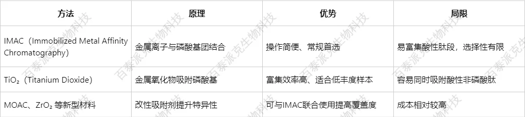
(2) Comparison of Mainstream Enrichment Methods:
4. Step Four: Sample Purification and Pre-treatment Before Loading
(1) Objective: To remove salts, buffers, and impurities, and to adapt to LC-MS platform requirements.
(2) Common Methods:
① C18 Solid Phase Extraction (SPE) column desalting;
② SpeedVac concentration and quantification;
③ Re-dissolve in a solution containing 0.1% FA to facilitate chromatographic separation.
5. Step Five: Mass Spectrometry Detection and Data Analysis
Mass spectrometry platform selection, parameter settings, database search, and statistical processing also affect the depth and credibility of final data.
III. Additional Suggestions for Improving Phosphoproteomics Data Quality
1. Reasonable Design of Experimental Groups and Replicates
(1) It is recommended to set ≥3 biological replicates per group to improve statistical confidence.
(2) When conducting quantitative experiments (such as TMT labeling), pay attention to batch effect control.
2. Focus on Site-Level Quantification Rather Than Protein-Level
Phosphorylation modifications have high site specificity, and biological interpretation should be combined with site quantification.
3. Utilize Bioinformatics Mining to Enhance Data Value
(1) Use methods such as motif analysis, pathway enrichment, and kinase prediction to explore potential regulatory networks.
(2) Recommended tools include: PhosphoSitePlus, KEGG, KinaseSubstrateDB.
The mass spectrometry analysis of phosphorylated proteins is a key means of revealing cellular signal networks, and high-quality sample preparation is the foundation of this process. If you encounter technical difficulties in actual operations or wish to apply phosphoproteomics to disease research, drug development, etc., feel free to contact Biotai Parker Biotechnology, and we will tailor solutions for you.
Biotai Parker Biotechnology—Characterization of Bioproducts, High-Quality Multiomics Mass Spectrometry Detection Service Provider
Related Services:
How to order?