


Analysis of Characteristic Peaks of Lysine in Circular Dichroism
Circular Dichroism (CD) is a technique used to study the secondary and tertiary structures of proteins, nucleic acids, and other biomolecules. By measuring the optical activity of molecules at specific wavelengths, information about their structures can be obtained.
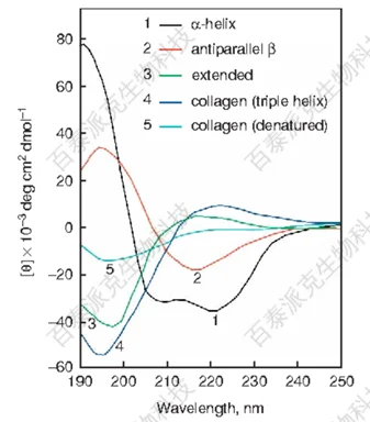
Figure 1
The optical activity of lysine (Lys) itself mainly appears in the far-ultraviolet region of the circular dichroism spectrum. For individual lysine or simple lysine peptides, characteristic peaks mainly appear in the far ultraviolet region (approximately 190-240 nm). In this region, the primary CD signal of lysine originates from its ε-amino group on the side chain, and these signals are often mixed with the CD signals generated by the protein backbone structure. Therefore, in large protein structures, the optical activity signals from lysine residues may be obscured by the background signals of the protein structure.
The CD characteristic peaks of proteins are mainly determined by their secondary structures (such as α-helix, β-sheet, random coil, etc.). For the secondary structure of proteins, here are some common CD characteristic peaks:
1. α-helix:
There is a positive characteristic peak at approximately 190-195 nm and two negative characteristic peaks at 208 and 222 nm.
2. β-sheet:
There is a positive characteristic peak at approximately 195 nm and a negative characteristic peak at approximately 215-218 nm.
3. Random coil:
Mainly exhibits a negative characteristic peak around 200 nm.
It should be noted that these wavelengths are approximate values, and the actual positions of characteristic peaks may vary depending on the specific sequence and conditions of the protein.
BiotechPack, A Biopharmaceutical Characterization and Multi-Omics Mass Spectrometry (MS) Services Provider
Related Services:
Circular Dichroism Analysis (CD)
Protein Structure Identification
How to order?
